Vulvar Cancer
A cancer affecting the external female genital area (vulva), often developing slowly and presenting with symptoms like itching, lumps, or abnormal skin changes.
Vulvar Cancer
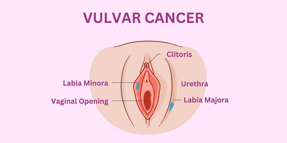
The vulva is the external part of the female genitals, including the clitoris, the vaginal lips, the opening to the vagina, and the surrounding skin and tissue. Most vulvar cancers are squamous cell carcinoma, which begins in thin, flat skin cells and is usually found on the vaginal lips.
A small number of vulvar cancers are adenocarcinomas, which begin in cells that produce mucus and other fluids. This type is usually found on the sides of the vaginal opening.
Vulvar cancer usually develops slowly over several years. Abnormal cells may grow on the surface of the vulvar skin for a long time, a condition known as vulvar intraepithelial neoplasia (VIN). Since VIN can progress to vulvar cancer, early detection and treatment are important.
Symptoms of Vulvar Cancer
Women with vulvar cancer may experience the following symptoms or signs. In some cases, there may be no symptoms.
Common symptoms include:
A lump or growth in or on the vulvar area
A patch of skin that is differently textured or colored than the surrounding area
Persistent itching, pain, soreness, or burning in the vulvar area
Painful urination
Bleeding or discharge that is not menstrual blood
An ulcer that persists for more than 1 month
A change in the appearance of an existing mole (especially in vulvar melanoma)
Wart-like growths similar to genital warts
These symptoms may be caused by cancer or other health conditions. It is important to consult a doctor if any of these symptoms occur.
Diagnosis of Vulvar Cancer
Local physical examination is the first step in diagnosing vulvar cancer. During the examination, the doctor inspects the vulva and evaluates the uterus, vagina, ovaries, fallopian tubes, bladder, and rectum for any abnormalities.
In some cases, swelling in the groin may be present, and a biopsy of the lymph node may be advised. This biopsy can be radical or sentinel.
A vulvar biopsy is performed to confirm the type of cancer and to help determine the appropriate treatment plan.
Surgical Treatment Options
Different vulvectomy approaches are used to treat invasive vulvar cancer:
Radical Local Excision of the Vulva:
Removal of the tumor along with a margin of surrounding tissue. It is used for most primary tumors less than 4 cm in size, typically in stage I or II disease.Modified Radical Vulvectomy:
A procedure where less than the full vulva is removed. For example, in radical hemivulvectomy, only one side of the vulva is removed.Radical Vulvectomy:
Removal of part or all of the vulva along with underlying deep tissue. This is less commonly performed, as many cases are managed with modified procedures or chemoradiation for larger tumors.Laser Surgery:
Uses a focused beam of light to remove premalignant lesions. It is not used for invasive tumors.Lymphadenectomy:
Surgical removal of lymph nodes in the groin to check for the spread of cancer.